


Scientists from the SLAC National Accelerator Laboratory (USA) have unveiled a method that significantly enhances the precision of sample preparation for cryoelectron tomography (cryo-ET). This breakthrough technique captures three-dimensional images of cellular internal structures with near-atomic resolution. The process, however, necessitates extremely thin samples, approximately 200 nanometers thick. Most cells, including human cells, pose a challenge as they are too “thick” for direct electron transparency. Researchers tackle this obstacle by employing an ion beam to excise thin slices from cells. Previously, achieving such precision required numerous attempts.
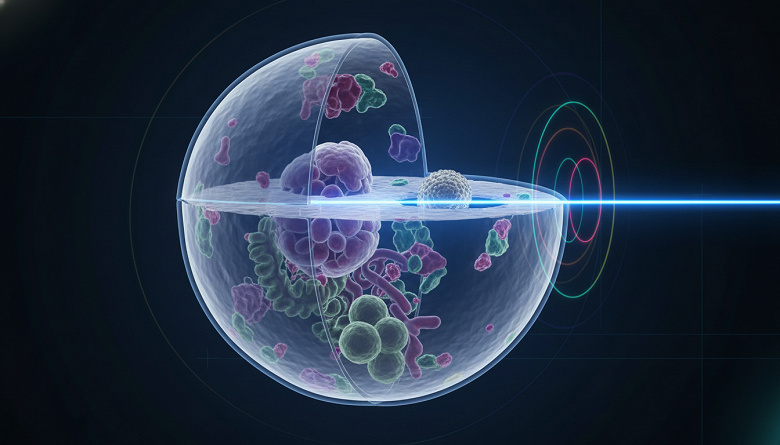
The novel method combines light microscopy with ion beam milling. Scientists utilize a tri-coincidence system aligning the focal planes of the scanning electron microscope, ion beam, and optical microscope. Unlike commercial systems, this setup allows for fluorescence tracking during milling. The principle is based on light interference-when the ion beam shears off the cell’s top, fluorescent light from an object tagged with special chemicals reflects and interferes with incoming light. Software developed for this method precisely determines the fluorescent object’s position based on brightness changes, enhancing the ion beam’s targeting accuracy.
Demonstrating the new method’s potential, researchers imaged a virus measuring 26 nanometers wide. The findings suggest this approach can target biological structures previously inaccessible to cryo-ET. The method may extend to studying other viral particles and small structures involved in cell division. In the future, the team plans to integrate advanced light microscopy techniques into the tri-coincidence system, further enhancing optical image quality and yielding maximally informative images for cryo-ET.
Since its introduction, other laboratories have begun exploring this method’s applications. Notably, the intricate alignment of microscopy systems could provide unprecedented insights into cellular mechanisms. The potential for refined data retrieval may revolutionize research in cell division and viral studies, offering detailed resolution that was previously unattainable. Employing advanced microscopy techniques can improve structural imaging and aid in analyzing complex biological interactions in various research fields.