


Breakthrough in Viewing Synaptic Activity
For the first time, a research team led by Professor Christian Rosenmund from Charite – Universitatsmedizin Berlin captured microscopic images documenting the release of neurotransmitters into the synaptic cleft. By utilizing optogenetically modified mouse neurons, which were stimulated by a light flash, the scientists triggered the secretion process. They instantly froze the cells in liquid ethane at a temperature of -180 °C just 1-2 milliseconds after the light pulse. This ‘instantaneous freeze’ allowed for visualization of structures using cryo-electron microscopy.
Adding to the significance, recent advancements in cryo-electron microscopy have refined our understanding of cellular processes at unprecedented resolutions, making it a cornerstone tool for neuroscience. This technique bridges gaps in our knowledge, providing tangible insights into molecular mechanisms.
Revealing the Synaptic Vesicle Fusion Process
Analysis of images revealed that the fusion of synaptic vesicles initiates with the formation of point junctions that subsequently expand into pores, allowing neurotransmitters to enter the synaptic cleft. Additionally, most fusing vesicles were linked by thin filaments to at least one other vesicle, which, according to researchers, ensures prolonged signal transmission.
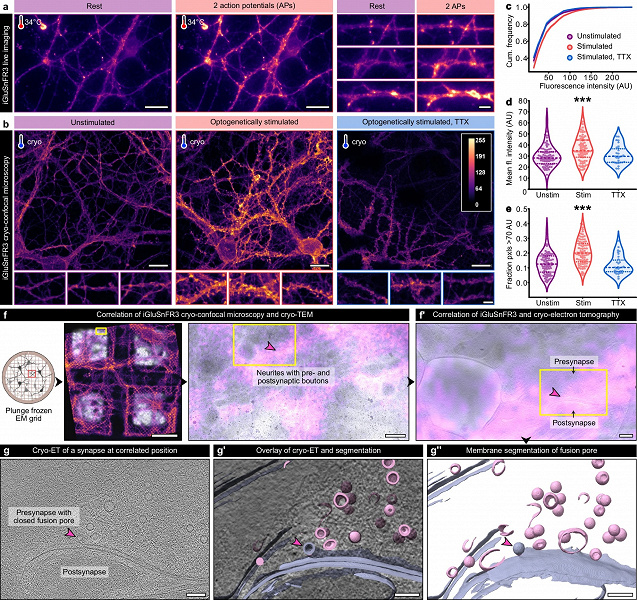
a – Electrical stimulation and live imaging of the glutamate sensor iGluSnFR3 at near-physiological temperatures: the left shows the state before stimulation, the right after (scale: overview 20 μm, enlarged fragments 10 μm). b – Maximum projections of neuron images without stimulation, after optogenetic stimulation, and after stimulation in the presence of tetrodotoxin (TTX), which blocks neuronal activity (scale: top images 20 μm, bottom 5 μm). c–e – Quantitative analysis of iGluSnFR3 fluorescence in individual axons: total intensity (c), average intensity (d), and the proportion of high-intensity pixels above 70 arbitrary units (e); after stimulation, the signal increases, and with the addition of TTX, this effect is suppressed; dashed lines indicate the median, dots – the 25th and 75th percentiles; differences are statistically significant (d: p)
“Until now, no one knew the exact stages of synaptic vesicle fusion with the cell membrane,” notes Dr. Jana Kroll, the study’s lead author working at the Max Delbruck Center. The developed technology has allowed synapses to be observed in action without disrupting their function.
Clinical Implications and Future Research
Understanding the detailed process of synaptic vesicle fusion, occurring millions of times per minute in the human brain, holds significant clinical importance. Mutations in proteins involved in this process are often found in individuals with epilepsy and other synaptic disorders.
“If we can identify the precise role of these proteins, it will be easier to develop targeted treatments for these so-called synaptopathies,” explains Rosenmund.
Dr. Kroll plans to repeat the experiments using human neurons derived from stem cells to better comprehend the differences in synaptic transmission mechanisms between mice and humans. Such endeavors hold promise for advancing personalized medicine, potentially leading to breakthroughs in treating neurological conditions linked to faulty neurotransmission.